Friday 13 May 2011

What is a gene mutation and how do mutations occur?

A gene mutation is a permanent change in the DNA sequence that makes up a gene. Mutations range in size from a single DNA building block (DNA base) to a large segment of a chromosome.
Gene mutations occur in two ways: they can be inherited from a parent or acquired during a person’s lifetime. Mutations that are passed from parent to child are called hereditary mutations or germline mutations (because they are present in the egg and sperm cells, which are also called germ cells). This type of mutation is present throughout a person’s life in virtually every cell in the body.
Mutations that occur only in an egg or sperm cell, or those that occur just after fertilization, are called new (de novo) mutations. De novo mutations may explain genetic disorders in which an affected child has a mutation in every cell, but has no family history of the disorder.
Acquired (or somatic) mutations occur in the DNA of individual cells at some time during a person’s life. These changes can be caused by environmental factors such as ultraviolet radiation from the sun, or can occur if a mistake is made as DNA copies itself during cell division. Acquired mutations in somatic cells (cells other than sperm and egg cells) cannot be passed on to the next generation.
Mutations may also occur in a single cell within an early embryo. As all the cells divide during growth and development, the individual will have some cells with the mutation and some cells without the genetic change. This situation is called mosaicism.
Some genetic changes are very rare; others are common in the population. Genetic changes that occur in more than 1 percent of the population are called polymorphisms. They are common enough to be considered a normal variation in the DNA. Polymorphisms are responsible for many of the normal differences between people such as eye color, hair color, and blood type. Although many polymorphisms have no negative effects on a person’s health, some of these variations may influence the risk of developing certain disorders.

What is mitochondrial DNA?

Although most DNA is packaged in chromosomes within the nucleus, mitochondria also have a small amount of their own DNA. This genetic material is known as  or mtDNA.
Mitochondria  are structures within cells that convert the energy from food into a form that cells can use. Each cell contains hundreds to thousands of mitochondria, which are located in the fluid that surrounds the nucleus (the cytoplasm).
Mitochondria produce energy through a process called oxidative phosphorylation. This process uses oxygen and simple sugars to create adenosine triphosphate (ATP), the cell’s main energy source. A set of enzyme complexes, designated as complexes I-V, carry out oxidative phosphorylation within mitochondria.
In addition to energy production, mitochondria play a role in several other cellular activities. For example, mitochondria help regulate the self-destruction of cells (apoptosis). They are also necessary for the production of substances such as cholesterol and heme (a component of hemoglobin, the molecule that carries oxygen in the blood).
Mitochondrial DNA contains 37 genes, all of which are essential for normal mitochondrial function. Thirteen of these genes provide instructions for making enzymes involved in oxidative phosphorylation. The remaining genes provide instructions for making molecules called transfer RNAs (tRNAs) and ribosomal RNAs (rRNAs), which are chemical cousins of DNA. These types of RNA help assemble protein building blocks (amino acids) into functioning proteins.

What is DNA?


DNA, or deoxyribonucleic acid, is the hereditary material in humans and almost all other organisms. Nearly every cell in a person’s body has the same DNA. Most DNA is located in the cell nucleus (where it is called nuclear DNA), but a small amount of DNA can also be found in the mitochondria (where it is called  or mtDNA).
The information in DNA is stored as a code made up of four chemical bases: adenine (A), guanine (G), cytosine (C), and thymine (T). Human DNA consists of about 3 billion bases, and more than 99 percent of those bases are the same in all people. The order, or sequence, of these bases determines the information available for building and maintaining an organism, similar to the way in which letters of the alphabet appear in a certain order to form words and sentences.
DNA bases pair up with each other, A with T and C with G, to form units called base pairs. Each base is also attached to a sugar molecule and a phosphate molecule. Together, a base, sugar, and phosphate are called a nucleotide. Nucleotides are arranged in two long strands that form a spiral called a double helix. The structure of the double helix is somewhat like a ladder, with the base pairs forming the ladder’s rungs and the sugar and phosphate molecules forming the vertical sidepieces of the ladder.
An important property of DNA is that it can replicate, or make copies of itself. Each strand of DNA in the double helix can serve as a pattern for duplicating the sequence of bases. This is critical when cells divide because each new cell needs to have an exact copy of the DNA present in the old cell.

Swine Flu

Swine influenza also known as Swine flu and carries the scientific name H1N1 influenza A and refers to influenza caused by any strain of the influenza virus found in pigs.
Swine flu is normally rare in humans. These strains infrequently circulate between humans and rarely mutates into a form able to pass from human to human. In humans, the symptoms of swine flu are similar to those of the common flu and of influenza-like illness and consist of namely chills, fever, sore throat, muscle pains, severe headache, coughing, and, weakness.
The 2009 outbreak in humans apparently is not due to a swine influenza virus. The strain is in most cases causing only mild symptoms and the infected people make a full recovery without medical attention and the use of antiviral medicines.

Paternity Testing

A paternity test is established to determine the biological father of a child like wise a maternity test is to prove the birth mother of a child. For obvious reasons paternity tests are much more common than maternity tests.
In a DNA paternity test, DNA samples from two possible fathers and the mother are compared with the offspring's DNA. In this procedure, the samples are digested with a type of enzyme that cuts DNA at specific sequences. The digested DNA is loaded onto a gel and separated according to size, by gel electrophoresis.
Every band of the offspring's DNA must match a band in at least one of its parents'. Let's first consider the offspring matches with the mother. Three bands match the mother. The three remaining bands must be shared with the father. Only one of the possible fathers will share the three bands with the offspring. Therefore, this must be the biological father.
Genetic testing has a 99.999% accuracy rate, or 99,999 out of 100,000 and has all but taken over the other forms of testing. Although there are several other, although often deemed less reliable parental tests. You could use several congenital traits sich as a cleft chin, a widows peak, or attached earlobes. Blood types can be used to disprove paternity in some cases. When the possible parents blood types are obtained this information can be used to show the possible blood types for the child, and because only certain blood type combinations are possible a mismatch may prove that the alleged mother or father of the child could not be the parent.

Speed of DNA Replication

The Genome of complex eukaryotes is huge and the process of DNA Replication should be incredibly fast. It is amazing that a Chromosome of 250 million pair of bases can be replicated in several hours. The speed of DNA replication for the humans is about 50 nucleotides per second per replication fork (low speed comparing to the speed of the bacterial DNA Replication).But the human Genome can be copied only in a few hours because because many replication forks take place at the some time (multiple initiation sites).

Enzymes of DNA Replication

Helicase: Unwounds a portion of the DNA Double Helix

RNA Primase: Attaches RNA primers to the replicating strands.

DNA Polymerase delta (ä): Binds to the 5' - 3' strand in order to bring nucleotides and create the daughter leading strand.

DNA Polymerase epsilon (å): Binds to the 3' - 5' strand in order to create discontinuous segments starting from different RNA primers.

Exonuclease (DNA Polymerase I): Finds and removes the RNA Primers

DNA Ligase: Adds phosphate in the remaining gaps of the phosphate - sugar backbone

Nucleases: Remove wrong nucleotides from the daughter strand.

Steps of DNA Replication


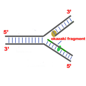
1)The first major step for the DNA Replication to take place is the breaking of hydrogen bonds between bases of the two antiparallel strands. The unwounding of the two strands is the starting point. The splitting happens in places of the chains which are rich in A-T. That is because there are only two bonds between Adenine and Thymine (there are three hydrogen bonds between Cytosine and Guanine). Helicase is the enzyme that splits the two strands. The initiation point where the splitting starts is called "origin of replication".The structure that is created is known as "Replication Fork"
.
2) One of the most important steps of DNA Replication is the binding of RNA Primase in the the initiation point of the 3'-5' parent chain. RNA Primase can attract RNA nucleotides which bind to the DNA nucleotides of the 3'-5' strand due to the hydrogen bonds between the bases. RNA nucleotides are the primers (starters) for the binding of DNA nucleotides.


3) The elongation process is different for the 5'-3' and 3'-5' template. a)5'-3' Template: The 3'-5' proceeding daughter strand -that uses a 5'-3' template- is called leading strand because DNA Polymerase ä can "read" the template and continuously adds nucleotides (complementary to the nucleotides of the template, for example Adenine opposite to Thymine etc).
b)3'-5'Template: The 3'-5' template cannot be "read" by DNA Polymerase ä. The replication of this template is complicated and the new strand is called lagging strand. In the lagging strand the RNA Primase adds more RNA Primers. DNA polymerase å reads the template and lengthens the bursts. The gap between two RNA primers is called "Okazaki Fragments".

The RNA Primers are necessary for DNA Polymerase å to bind Nucleotides to the 3' end of them. The daughter strand is elongated with the binding of more DNA nucleotides.

DNA Pol I - exonuclease

DNA Double Helix


Lets have a look in how the DNA Double Helix (two DNA strands) looks like. Every DNA strand is consisted of nucleotides. Nucleotides are monomers that are made of a phosphate, a sugar (deoxyribose) and a heterocyclic base (Thimine, Cytosine -pyrimidines- Adenine, Guanine - purines-). The combination of a sugar and an heterocyclic base gives a nucleoside. When a phosphate is added to the molecule, a nucleotide is created.
The backbone of each strand consists of alternating phosphates and deoxyriboses. To be more specific, the phosphate of a nucleotide bonds both to the 5' carbon of one deoxyribose and the 3' carbon of the next deoxyribose. This is how the nucleotides create the strand. The 5' and 3' refers to the place of the carbons in the deoxyribose molecule. The heterocyclic bases which are attached to the deoxyriboses project in towards the axis of the helix.

The two polynucleotide strands wound around each other. The strands of a DNA double Helix are antiparallel which means that the one chain runs 5'-3' and the other runs 3'- 5'. The connections of the two strands are due to the hydrogen bonds between the nucleotide bases of the two strands. Connections happen between the adenines and thymines (2 hydrogen bonds) and between cytosines and guanines (3 hydrogen bonds). The two strands are complementary, which means that the strand running in the 5'A 3' direction will have base Adenine that will pair with base Thymine on the opposite strand running in 3'A 5' direction. Since the strands are complementary and antiparallel, each of them can be a matrix for the creation of a new complementary and antiparallel strand, as we can see in another chapter of this site.